
Our Products
Rayland Medical's consistent self produced and selected products are guaranteed to provide safety for you and your customers
Our Products
Portable and handheld ultrasound systems have improved rapidly, but compact size does not eliminate the technical trade-offs involved in ultrasound imaging.
For procurement teams, “HD imaging” or “high image quality” is too broad to compare products effectively. Image quality depends on probe frequency, penetration depth, number of elements, beamforming, display, frame rate, Doppler sensitivity, patient anatomy, presets, and operator experience.
Understanding where portable ultrasound is most likely to reach its limits helps avoid purchasing a device that works well in a demonstration but struggles with the examinations it was actually purchased to perform.
One important distinction is between image quality and clinical usefulness.
A randomized comparison of handheld and cart-based ultrasound found that the cart-based equipment produced better overall image quality, but diagnostic accuracy was similar for the studied cardiac, lung, biliary, renal, and abdominal-aorta examinations.
Another comparison of handheld and traditional ultrasound for RUSH examinations found adequate images for interpretation in 82% and 86% of examinations respectively, without a statistically significant difference between the devices.
This means portable ultrasound may be entirely suitable for a focused clinical question even when a premium cart system produces a more refined image.
Ultrasound frequency creates a fundamental trade-off.
Higher frequencies provide better resolution but do not penetrate as deeply. Lower frequencies penetrate further but provide less fine detail.
This is why linear probes are normally used for superficial structures, while lower-frequency convex or phased-array probes are preferred for deeper abdominal and cardiac imaging.
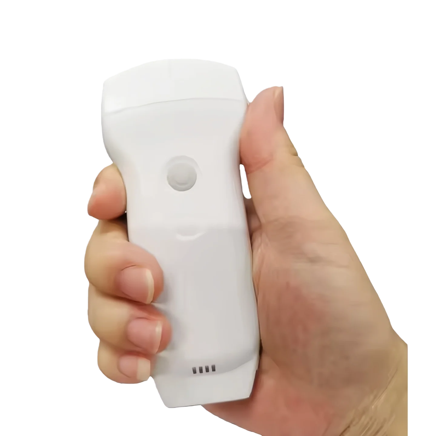
Rayland's dual handheld scanner addresses these different requirements with a linear side listed at 7.5/10 MHz and a convex side with lower-frequency settings for abdominal and deeper examinations.
For larger patients or very deep targets, however, image quality should be checked with actual clinical samples rather than assuming that a listed maximum depth guarantees sufficient diagnostic detail.
Portable systems may have more difficulty displaying very small structures or subtle tissue differences.
In one musculoskeletal comparison, portable ultrasound missed some small findings and showed lower color-Doppler sensitivity than the cart-based system.
This matters when the target includes:
A handheld scanner intended mainly for vascular access or bedside screening has a different image-quality requirement from a system intended to make detailed radiological diagnoses.
B-mode image quality is only one part of the comparison.
A system may produce a clear grayscale image but have weaker detection of slow blood flow.
For vascular, inflammatory, cardiac, or lesion assessment, check:
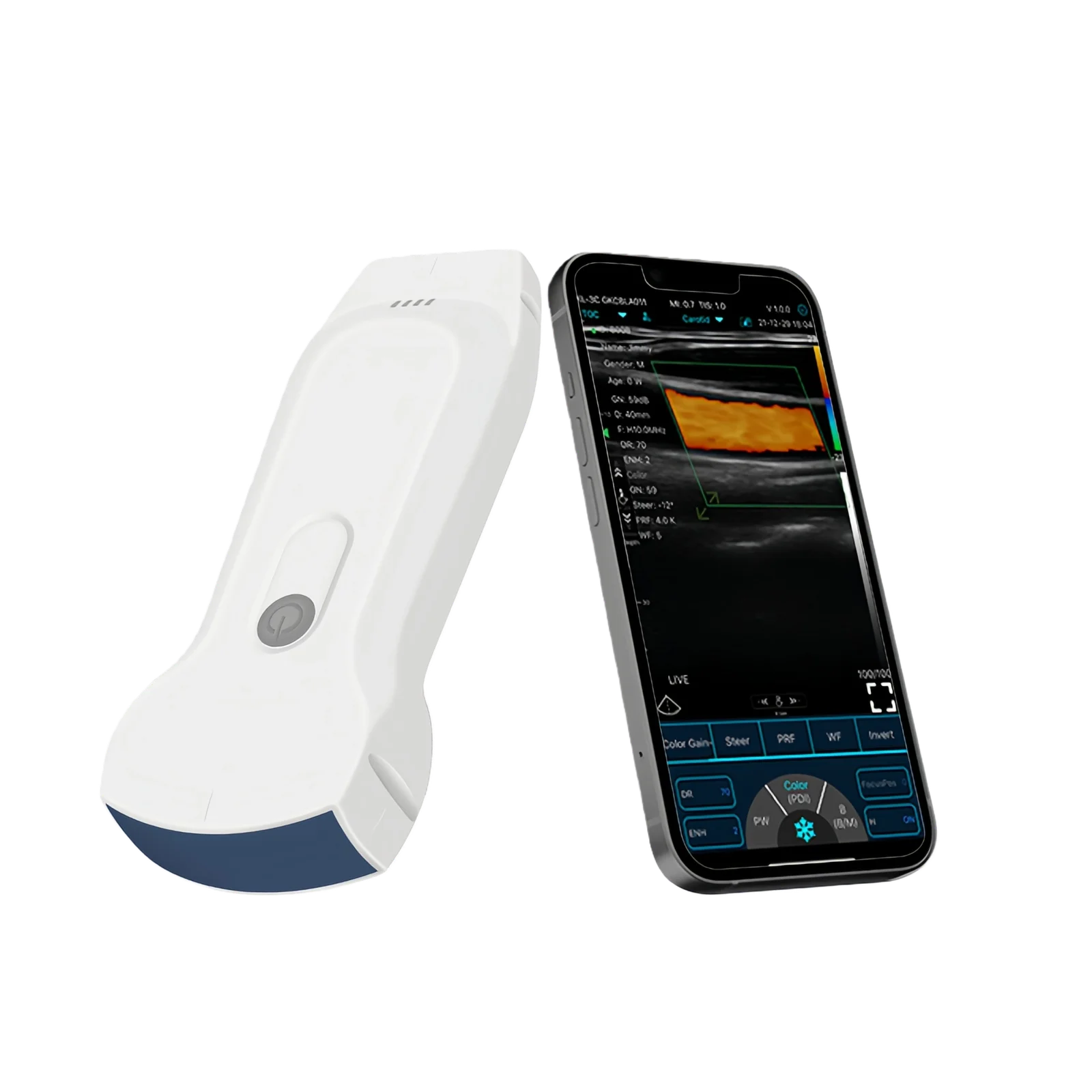
Rayland's handheld ultrasound currently provides Color, PDI, and PW modes as well as automatic vessel-flow measurements.
If Doppler is a major purchasing requirement, suppliers should provide sample images or demonstrations from the actual application rather than relying on the presence of “Color Doppler” in the specification table.
Frame rate becomes particularly noticeable when observing moving anatomy such as the heart.
Rayland's current handheld system specifies an image frame rate of 24 frames per second.
Whether that is sufficient depends on the intended examination.
For basic cardiac POCUS, the priority may be identifying gross ventricular function, pericardial fluid, chamber size, or other focused findings.
Detailed echocardiography may demand more sophisticated cardiac imaging capabilities.
The procurement question should therefore be what cardiac decision must the image support?
Portable ultrasound is often displayed on a smartphone or tablet.
Even when the probe captures adequate data, a small screen can make subtle findings harder to evaluate.
Screen quality depends on:
A smartphone may be ideal for ambulance or bedside use. A larger tablet may be preferable for routine clinical interpretation.
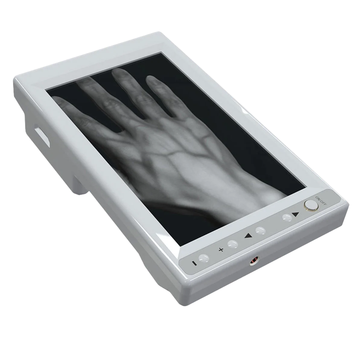
If an OEM handheld ultrasound project will be supplied together with a dedicated tablet, the display should be tested as part of the complete system.
Different examinations require different combinations of depth, gain, frequency, focus, Doppler settings, and image processing.
Poor presets can make capable hardware look weak.
Rayland's dual-probe system currently includes presets for abdomen, kidney, urology, gynecology, obstetrics, lung, cardiac, vascular, thyroid, breast, small parts, MSK, nerve, and other applications.
For distributors evaluating an OEM product, preset quality should therefore be tested for the target departments.
A supplier demonstration using only one abdominal image does not prove that thyroid, vascular, cardiac, and MSK presets are equally well optimized.
Image quality should be tested on more than ideal subjects.
Greater tissue depth increases attenuation and can make ultrasound imaging more challenging. Rib spaces, bowel gas, wounds, dressings, body habitus, and patient positioning may also restrict the acoustic window.
ACEP equipment-selection guidance specifically recommends testing image quality under challenging patient conditions rather than evaluating only easy scans.
For procurement, ask for evaluation in the hardest common use case—not the easiest.
A poor image is not always an equipment problem.
Probe angle, contact, gain, depth, focus, preset selection, and anatomical knowledge all affect image acquisition.
The same portable system can therefore produce very different results in the hands of an experienced POCUS operator and a first-time user.
Training should be included in the implementation plan, particularly where handheld ultrasound is being introduced into departments that previously relied on radiology staff.
Rather than requesting one attractive sample image, prepare several typical examinations.
For example:
Convex probe: abdomen, kidney and bladder
Linear probe: vessel, thyroid and superficial MSK
Cardiac preset: basic cardiac views
Doppler: vascular flow
Compare:
Rayland Medical's handheld system combines convex and linear scanning in a single portable device with 128 elements, multiple imaging modes, examination presets, and mobile-device connectivity.
Portable ultrasound should not be expected to outperform every high-end cart system. The relevant question is whether its image quality is sufficient and repeatable for the examinations included in the purchasing plan.