
Our Products
Rayland Medical's consistent self produced and selected products are guaranteed to provide safety for you and your customers
Our Products
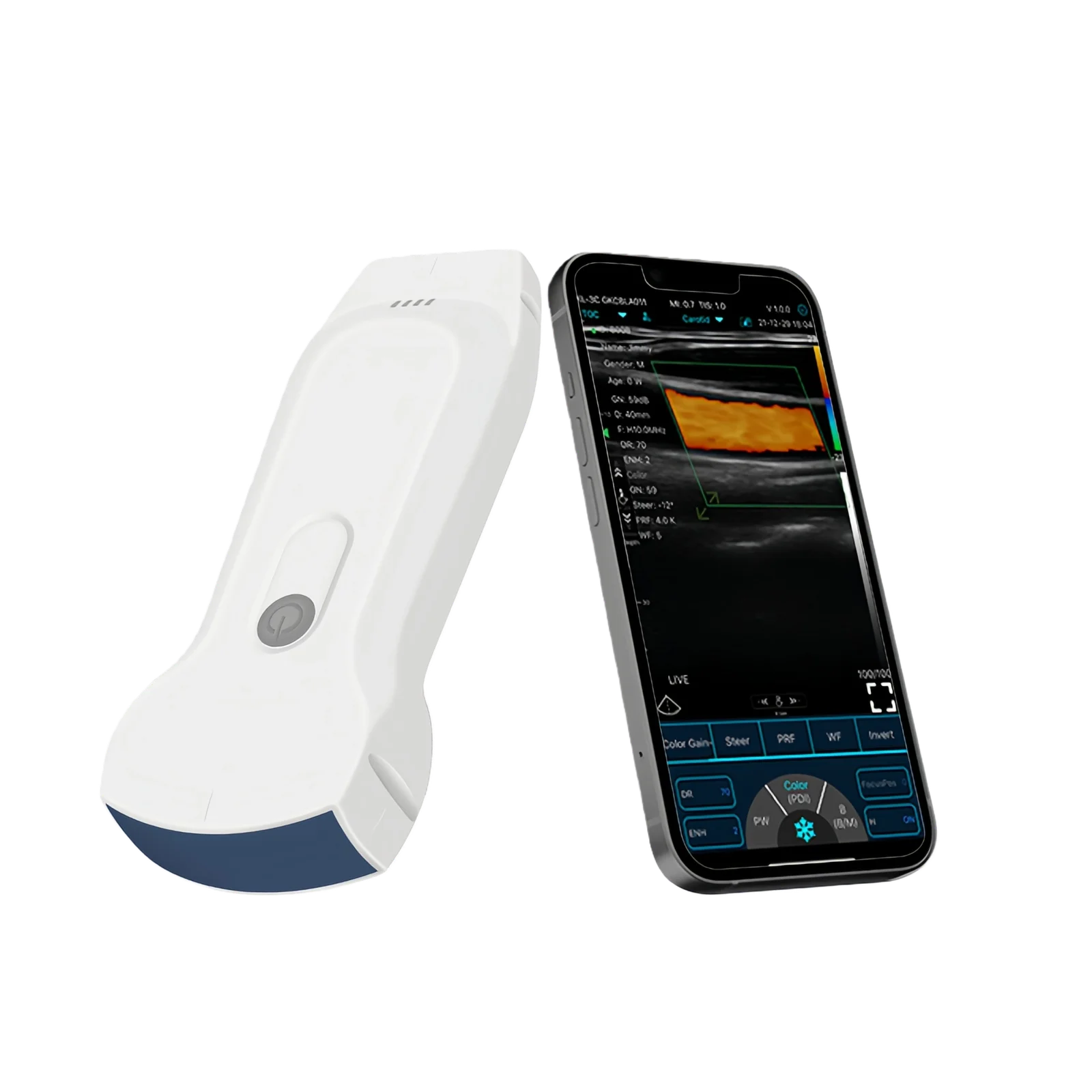
Handheld ultrasound has already solved much of the hardware portability problem. A probe can connect to a smartphone or tablet and provide bedside imaging without moving a conventional ultrasound cart.
The next challenge is the operator.
Ultrasound remains highly dependent on how the clinician positions the probe, recognizes the correct view, adjusts the image, makes measurements, and interprets the result.
This is where artificial intelligence is beginning to change point-of-care ultrasound.
AI-enabled systems can assist with image acquisition, image-quality assessment, measurements, anatomy recognition, and selected interpretation tasks. But these capabilities should be evaluated individually rather than treating “AI ultrasound” as one single feature.
A handheld scanner can be physically available to every department, but that does not mean every clinician can immediately acquire a diagnostic image.
The user must know:
A 2026 review of AI-assisted POCUS described this as a growing mismatch between widely available handheld hardware and limited expert mentorship. The review found that AI-guided probe positioning, view recognition, anatomy labeling, automated capture, and quality assessment may help novice users acquire adequate images for selected applications, although broader validation is still needed.
One emerging function is real-time acquisition guidance.
The software analyzes the current image and provides instructions such as:
This is particularly relevant for cardiac POCUS because obtaining standard cardiac views can be difficult for inexperienced operators.
A randomized study involving novice clinicians found that users with deep-learning guidance acquired apical four-chamber cardiac images faster and achieved higher image-quality scores after a period of use than those using a non-AI handheld device.
A newer randomized trial evaluating multiple echocardiographic views likewise reported faster acquisition and better image-quality scores among novices using deep-learning guidance.
These findings are promising, but they apply to defined tasks and should not be generalized to every ultrasound examination.
Another useful function is automated image-quality assessment.
Instead of leaving a beginner uncertain whether the correct anatomy has been captured, an algorithm can assess whether the view meets predefined criteria.
This can potentially reduce repeated scanning and create more standardized datasets for later review.
For training programs, it may also provide immediate feedback between formal supervised sessions.
However, quality scoring is not the same as diagnosis.
A technically adequate image can still require expert clinical interpretation.
AI and advanced image processing can also automate selected measurements.
Examples across modern ultrasound platforms include automatic:
Automation can reduce manual caliper placement and improve workflow consistency.
But procurement teams need to distinguish between:
automatic calculation based on conventional image processing
and
AI-enabled clinical software validated for a specific intended use.
Not every “Auto” button represents artificial intelligence.
The main potential value is not replacing experienced sonographers or physicians.
It is lowering the acquisition barrier for defined bedside questions.
A recent review suggests that AI-supported acquisition may act as a type of “digital mentor,” allowing less experienced users to obtain acceptable images for focused examinations after appropriate training.
This could be useful in:
The benefit is greatest when the clinical question is narrow and well defined.
AI cannot recover anatomical information that the probe never captured.
Probe type, frequency, penetration, element count, frame rate, Doppler performance, and image quality still determine what information reaches the algorithm.
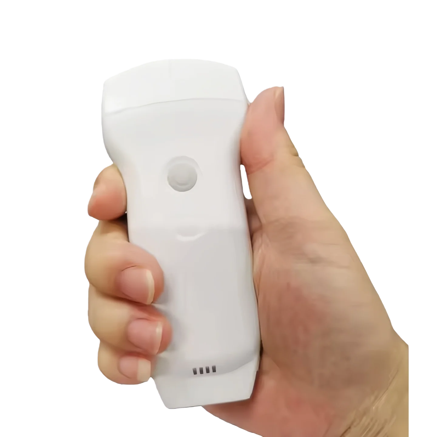
Rayland Medical's current handheld ultrasound provides a 128-element dual-probe architecture, with a convex array for deeper abdominal and selected cardiac imaging and a 7.5/10 MHz linear array for vascular, nerve, thyroid, MSK, and other superficial examinations. It supports USB/WiFi connection and multiple B-mode and Doppler functions.
This type of hardware platform determines the clinical imaging foundation on which any future advanced software functions must operate.
Current handheld scanners commonly provide examination presets.
Rayland's system includes presets for abdomen, urology, gynecology, kidney, obstetrics, lung, cardiac, thyroid, carotid, breast, pediatrics, MSK, vascular, nerve, and vessel flow.
A preset automatically configures parameters suitable for a particular examination.
That improves workflow, but it should not automatically be marketed as artificial intelligence.
For an AI-enabled OEM project, the supplier should state exactly:
Software that contributes to diagnosis or clinical decision-making can be part of a regulated medical device.
The FDA maintains a continuously updated list of AI-enabled medical devices that have met applicable US premarket requirements. Ultrasound systems and ultrasound-related AI products are already represented in that list.
Therefore, adding an AI function is not equivalent to adding another display color or language.
For OEM procurement, ask whether the AI-enabled configuration is covered by the product's existing regulatory authorization.
Some AI functions operate locally on the device.
Others may require:
That changes hospital IT requirements.
POCUS implementation guidance emphasizes security, patient confidentiality, image storage, archiving, and integration into clinical systems as part of responsible deployment.
Before purchasing AI-enabled handheld ultrasound, confirm where patient data is processed and stored.
This is one of the most important limitations.
An algorithm may help the user reach an appropriate image, but the clinician still needs to understand:
Current literature on AI-assisted POCUS repeatedly notes limitations involving external validation, different devices, different patient body habitus, algorithm generalizability, and clinical outcomes.