
Our Products
Rayland Medical's consistent self produced and selected products are guaranteed to provide safety for you and your customers
Our Products
Terms such as Standard Mode, Enhanced Mode, Fine Mode, and DVA Mode are increasingly used when describing infrared vein viewers.
However, these are not universally standardized medical-device mode names.
One manufacturer's “DVA Mode” may be another manufacturer's “Enhanced Mode,” “Fine Mode,” or simply an advanced image-processing setting.
For hospital procurement, it is therefore better to understand what the mode actually changes than to compare the name printed on the interface.
A standard mode normally provides general-purpose NIR vein visualization for routine patients.
The imaging pipeline typically:
A standard mode usually tries to balance:
This can work well when veins are already relatively superficial and the goal is simply to make their path easier to see.
A DVA-focused mode typically applies stronger processing to difficult vascular images.
Depending on the manufacturer, it may change:
The purpose is to make a low-contrast venous pattern more distinguishable.
That can be useful for patients with obesity, edema, small pediatric vessels, chemotherapy history, or otherwise difficult superficial vascular access.
However, stronger enhancement does not automatically mean that the displayed vessel is deeper or more suitable for cannulation.
This point matters when writing an RFQ.
Do not specify simply:
Must include DVA Mode.
Two manufacturers may interpret that requirement differently.
Instead, describe the performance you need, such as:
That allows meaningful comparison across suppliers.
Rayland Medical does not currently publish a simple “Standard Mode / DVA Mode” switch on the PRO 500/600 specification.
Instead, its DVA-oriented PRO platform combines multiple image-display options and processing functions.
The PRO 500 currently lists 10 display colors. The PRO 600 lists 16 display colors, digital color blending, and deep-neural-network computation. Both operate in an 850–940 nm wavelength range and are intended for DVA populations such as children, obese patients, patients with swelling, repeated chemotherapy, or poor vascular elasticity.
This illustrates why comparing underlying functionality is more useful than comparing mode names.
Increasing contrast makes vessels stand out.
Too much processing, however, can potentially make a vessel look wider, narrower, or more sharply defined than the raw optical signal would suggest.
The clinician should therefore retain the ability to compare different visual modes.
A useful DVA mode should improve recognition without making the display unstable or difficult to interpret.
During product evaluation, examine:
The best mode is not necessarily the one producing the darkest vessel image.
Changing display color is not the same as increasing vein-detection depth.
Colors are primarily used to improve visual contrast for the user under different patient and environmental conditions.
Rayland's vein-finder range provides several colors and inverse display configurations.
A procurement demonstration should allow users to test which colors remain easiest to interpret under normal treatment-room lighting.
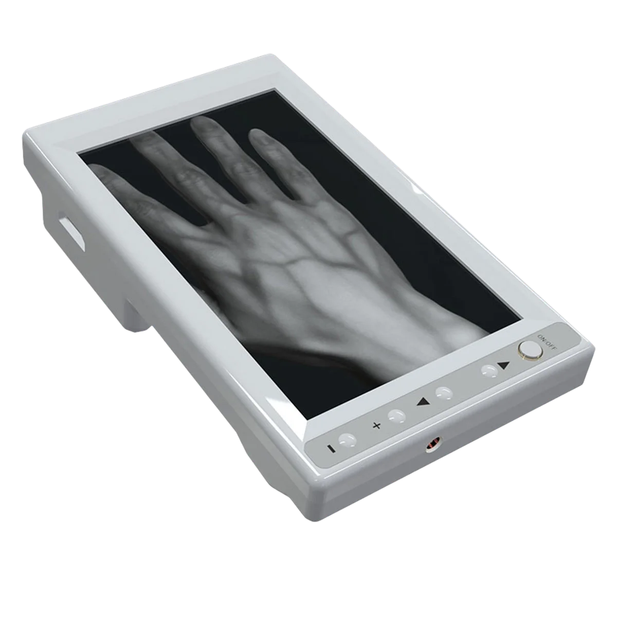
In normal display, veins may appear dark against a lighter background.
Inverse mode reverses this relationship.
Some users find one format easier for:
The clinical information has not changed; only the way it is presented has changed.
This is why multiple viewing options can be useful without implying that each mode represents a different clinical technology.
A common marketing misunderstanding is that a stronger or “deep” mode allows an NIR vein finder to perform the same job as ultrasound.
It does not.
NIR is still limited mainly to superficial vascular visualization.
If a patient's usable vessel is too deep to be visualized optically, ultrasound may be more appropriate because it can show the vessel in cross-section and provide depth, diameter, compressibility, surrounding anatomy, and real-time needle guidance.
For severe DVA, a hospital may therefore need both technologies at different stages of the vascular-access pathway.
Rayland's PRO 600 specification includes deep-neural-network computation.
For procurement, the relevant question is not:
“Does it use AI?”
It is:
“Does the processing produce a clearer and more accurately aligned vessel image in the patients we actually struggle with?”
Evaluate difficult cases rather than accepting an algorithm name as proof of performance.
Prepare several patient scenarios and compare:
Standard/easy access: Can the image remain natural and stable?
Small pediatric veins: Can small vessel paths be distinguished?
Obese patient: Does enhancement improve useful superficial visualization?
Darker skin tone: Which display color and contrast setting works best?
Edema: Can vessel structures be separated from background changes?
Also evaluate projection accuracy, processing latency, battery life, ergonomics, and available stands.
Rayland's vein-finder platform can be used handheld or with desktop and mobile support configurations, allowing the same imaging device to fit different clinical workflows.