
Our Products
Rayland Medical's consistent self produced and selected products are guaranteed to provide safety for you and your customers
Our Products
An infrared vein finder does not “see through” the human body in the same way as ultrasound or X-ray imaging.
Instead, it takes advantage of differences in how biological tissues absorb and reflect near-infrared light.
The device illuminates the skin, detects the reflected optical information, processes the difference between blood vessels and surrounding tissue, and then displays or projects a high-contrast representation of the superficial venous pattern.
Understanding this process makes it easier to compare vein finders by meaningful technical parameters rather than simply by brightness or the number of display colors.
Visible light is strongly affected by skin pigmentation and has limited tissue penetration.
Near-infrared wavelengths can penetrate several millimeters into tissue while still interacting with hemoglobin.
A review of vein-finder technologies found that wavelengths between approximately 740 and 940 nm are commonly used for peripheral vein visualization, with 850 nm being particularly common among investigated systems.
Rayland Medical's standard vein finder uses approximately 850 nm NIR light, while the PRO 500/600 specification lists an 850–940 nm wavelength range.
The exact wavelength is only one part of image performance. Sensor sensitivity, illumination uniformity, optical filters, software processing, and projection alignment also matter.
When near-infrared photons reach tissue, several things happen.
Some light is:
Blood and surrounding tissue do not return exactly the same optical signal.
Hemoglobin contributes to the absorption characteristics of blood, creating contrast that can be detected by an NIR-sensitive imaging system.
Research on vein visualization has shown that veins containing deoxygenated hemoglobin can form darker contrast against surrounding tissue because of differences in absorption and backscatter.
The device converts these subtle differences into an image that is much easier for the human eye to interpret.
The vein finder first directs near-infrared light onto the selected anatomical area.
Good illumination needs to be sufficiently uniform.
If one region receives much more light than another, the image-processing system has to compensate for uneven brightness before it can reliably distinguish vessel structures.
The working distance also matters.
Rayland's PRO models specify a detection distance of approximately 20–25 cm, while its WHITE model lists about 20 cm ±3 cm.
Maintaining the intended distance helps keep the projection scale and optical focus consistent.
The reflected and scattered light is collected by an imaging sensor.
The raw image is not necessarily useful to the clinician.
It may contain:
This is where image processing becomes important.
The system must separate useful vascular information from surrounding optical information quickly enough to produce a real-time image.
Image-processing algorithms can increase the difference between probable venous structures and surrounding tissue.
Depending on the platform, processing can include:
Rayland's PRO 600 lists digital color blending and deep-neural-network computation as part of its image-processing configuration.
These features should be evaluated according to the resulting image rather than simply treating “AI” or “neural network” as proof of better clinical performance.
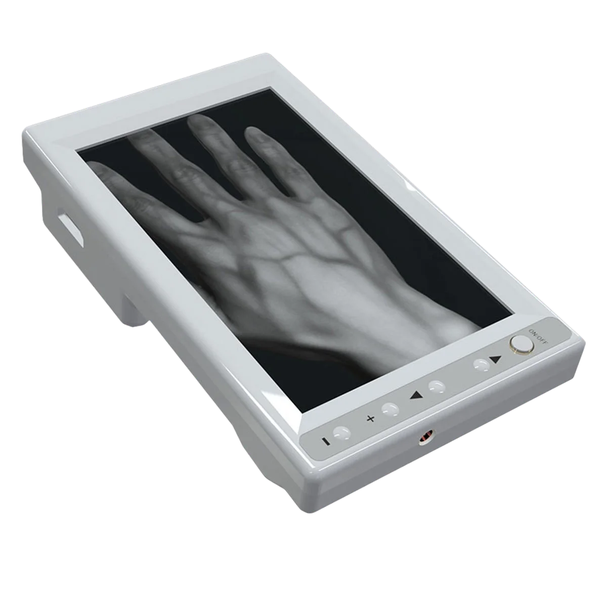
After processing, the vascular pattern can be projected onto the patient's skin or presented through the device's display system.
The objective is to align the visualized vein path with the underlying anatomy closely enough that the clinician can use the information when selecting the puncture site.
Projection accuracy is therefore one of the most important practical parameters.
A bright image that is displaced from the true vessel location can be less useful than a lower-brightness image with better alignment.
During product evaluation, clinicians should move the device around the working area and check whether the projected vascular path remains stable.
This depends on the display mode.
The raw optical information can be converted into many different visual presentations.
A device may show:
The underlying anatomy has not changed.
The system is simply remapping contrast so the user can select whichever presentation is easiest to recognize.
Rayland's vein-finder range includes multiple colors and inverted modes for this reason.
Melanin, tissue thickness, hair, subcutaneous fat, and ambient illumination can all influence the optical signal.
The advantage of NIR is that it can reduce some of the limitations of visible-light inspection, but it does not make patient-to-patient differences disappear.
Studies of NIR vein-finder design have reported that optimum wavelength and image quality can vary with skin color and tissue characteristics.
This is why adjustable contrast, color, and image-processing modes can matter in a hospital serving a diverse patient population.
NIR vein visualization is mainly a superficial vascular visualization technology.
A review of vein-finder prototypes reported tissue penetration on the order of several millimeters in the commonly used NIR wavelength range.
Actual useful visualization depth depends on device design and patient anatomy.
Procurement teams should be cautious when comparing manufacturers that publish a single maximum “vein depth” number without describing the test conditions.
Image clarity on a superficial hand vein does not prove equivalent performance on a deeper vein in an obese or edematous patient.
This distinction is essential.
NIR vein visualization produces an optical map of superficial vessels.
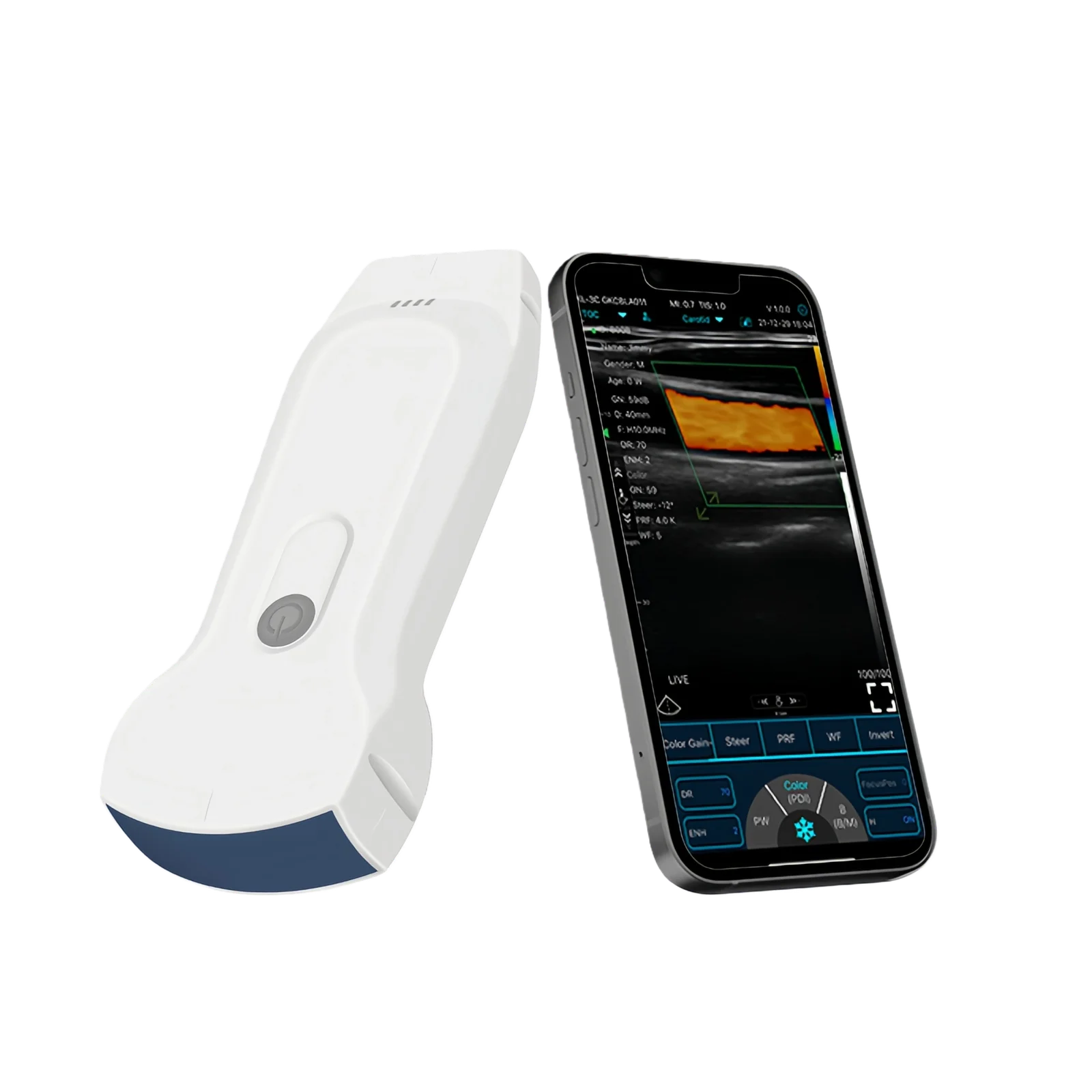
Ultrasound produces a cross-sectional image based on reflected sound waves.
Ultrasound can therefore provide information such as:
A vein finder is simpler and faster for superficial vein visualization, while ultrasound offers more information for deeper or more complex vascular access.
Wavelength alone does not determine performance.
For procurement, evaluate the complete imaging chain:
NIR Source → Optical Sensor → Image Processing → Projection → Alignment → User Interface
Useful comparison points include projection accuracy, small-vein visibility, latency, working distance, skin-tone performance, display modes, battery time, and mobility.
Rayland Medical's PRO 500/600 combines NIR imaging with multiple display colors, rechargeable battery operation, and handheld or support-mounted configurations.
The science behind the system is relatively straightforward. The engineering challenge is making the optical difference between veins and surrounding tissue clear, stable, accurately aligned, and useful in real time.